Embryology | IVF | Embryo Biopsy
Blastocyst Biopsy: Before and After
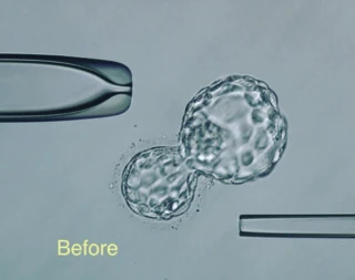
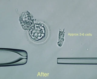
Here we see the before and after pictures of a biopsy done last week. In the first picture, you can see a beautiful 4Aa embryo. In the second, the 3-6 cells biopsied after the procedure.

 The biopsy itself takes about 30 seconds. However, with setup, tubing, and paperwork, each case is about 15 minutes.
In the after picture you can see the intact embryo showing no loss of cells (besides the obvious!) and it's actually still expanded slightly inside the zona. From here, your embryologist would collapse it completely with a quick laser shot and it would be vitrified shortly after.
Clinics take around 3-6 cells each time so that an accurate reading can be produced when genetic testing is performed. Taking too few cells may result in an inconclusive answer (not ideal - need to thaw and rebiopsy) and taking too many can harm the embryo! It's a fine balance between the two.
Of course, biopsy is not for the unskilled embryologist and more often than not it takes a great deal of practice to get reproducible results. Ideally, patients should ask labs what their post-thaw survival rates are following biopsy (meaning: how many embryos survive the biopsy and freeze) as this gives an indication of the skill of the operator.